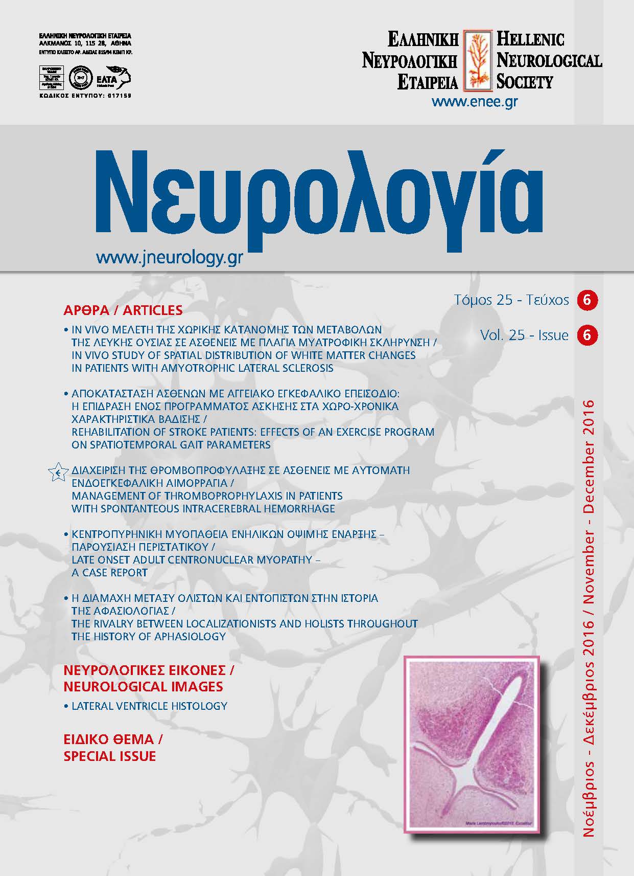
“EXCALIBUR” like Lateral Ventricle Histology
Abstract
Tissue section of fetal brain in which a big sword has been discovered in a hematoxylin and eosin
(H&E) slide after a hard day working on microscope.
The lateral ventricle is covered with ependyma and in this histological section seems like “Excalibur”. Ependyma is a continuous cuboidal or columnar epithelium that lines the brain ventricles and
the central canal of the spinal cord. These cells bear apical microvilli to increase surface area, and
most also have motile cilia that project into the ventricular lumen. Luminal surfaces of ependymal
cells are in direct contact with CSF. Another characteristic of the ependymal cells is the presence of
apical intracellular junctions in order to serve a protective and selective barrier between brain and
CSF and prevent passage of potentially neurotoxic substances to the brain.
The dense layer of small dark cells below the ependymal epithelium is the germinal matrix. This
histological structure is a highly cellular and highly vascularized region from which cells migrate out
during brain development, mainly between 22nd and 30th weeks of gestation.